
LEADTOOLS Medical SDK
Develop powerful medical imaging applications.
Published by LEADTOOLS
Distributed by ComponentSource since 1996
Prices from: US$ 8,085.00 Version: v23 Updated: Mar 5, 2024
Develop powerful medical imaging applications.
Published by LEADTOOLS
Distributed by ComponentSource since 1996
Prices from: US$ 8,085.00 Version: v23 Updated: Mar 5, 2024

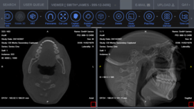
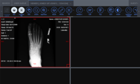
LEADTOOLS Medical SDK is ideal for developing medical applications. It features comprehensive DICOM data set support, 8-16 bit extended grayscale image support, image annotation, specialized extended grayscale image display such as window level and LUT processing, and medical-specific image processing. Other features include lossless JPEG compression, and signed and unsigned image data processing. LEADTOOLS Medical Imaging Suite builds on the functionality providing more advanced features, including HTML/JavaScript viewing.


Image 1 / 9
Dmitry Smirnov, Samsung Research Center
LEADTOOLS Medical SDK Includes:


LEADTOOLS Medical SDK is also available in:
Live Chat with our LEADTOOLS licensing specialists now.


Tel: (888) 850 9911
Fax: +1 770 250 6199


